Image Gallery Main Menu Yeasts Opportunistic Fungi Dematiaceous Fungi
Dimorphic Fungi Dermatophytes Actinomycetes Direct Microscopy
Pneumocystis carinii Technical Cases

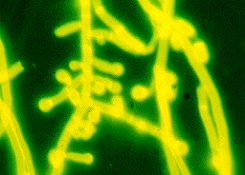
Microsporum persicolor

Stain: Calcofluor White

Magnification: x1000


Structure: Club shaped or round microconidia (often pedicellate); smooth walled, fusiform, bullet shaped macroconidia containing 4 to 7 cells. Frequent spiral hyphae are seen (not shown in this image).


© Copyright 1999-2007 Department of Microbiology, Mount Sinai Hospital, Toronto, Canada. All rights reserved.